Reproductive Health Sexual and Reproductive Health
Same Origin, Different Forms: Homologous Structures
Have you ever considered how profoundly similar the male and female body plans are? While we often focus on the differences, a closer look at reproductive anatomy reveals a fascinating story of shared origins. This story is told through homologous structures—body parts that arise from the same embryonic tissue but develop into different forms.
Understanding these structures is more than just a biological curiosity. It offers deep insights into human development, sexual differentiation, and even our evolutionary history. By exploring the common beginnings of male and female anatomy, we can better appreciate the intricate processes that shape us and the clinical significance when these processes vary. This post will guide you through the essentials of male and female homologous structures, from their embryonic origins to their fully formed functions.
What Are Homologous Structures?
In biology, homologous structures are organs or body parts in different organisms that share a common ancestry or developmental origin. A classic example is the forelimb of vertebrates: the wing of a bat, the flipper of a whale, the leg of a cat, and the arm of a human all share a similar underlying bone structure, despite their different functions. This similarity points to a shared evolutionary ancestor.
When we apply this concept to human anatomy, male and female homologous structures are parts of the reproductive system that originate from the same embryonic tissues. Early in development, the reproductive anatomy of an embryo is bipotential, meaning it has the potential to develop into either a male or female system. As development progresses, hormonal signals guide these tissues to differentiate, creating the distinct reproductive structures we recognize in males and females.
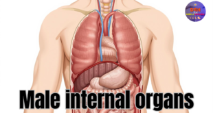
From Common Ground: Reproductive Systems
The internal reproductive organs in males and females provide a clear example of homology. They start from the same foundational tissues before specializing into their distinct roles.
Testes and Ovaries
The primary reproductive organs, or gonads, are the testes in males and the ovaries in females. Both develop from the same embryonic tissue called the gonadal ridge.
- Testes (Male): The testes are responsible for producing sperm and the primary male sex hormone, testosterone. They descend from the abdomen into the scrotum during fetal development or shortly after birth.
- Ovaries (Female): The ovaries produce eggs (ova) and the primary female sex hormones, estrogen and progesterone. They remain within the pelvic cavity, near the uterus.
Spermatic Cords and Suspensory Ligaments
The structures that support the gonads also share a common origin.
- Spermatic Cords (Male): These cords are bundles of nerves, ducts, and blood vessels that run from the abdomen down to the testicles. They are crucial for the function and health of the testes.
- Suspensory Ligament of the Ovary (Female): This ligament extends from the ovary to the wall of the pelvis. It contains the ovarian artery and vein, providing a vital blood supply to the ovary. Both the spermatic cord and the suspensory ligament are remnants of the gubernaculum, a structure that guides the descent of the gonads.
A Shared Blueprint: External Genitalia
The external genitalia offer some of the most striking examples of male and female homologous structures. These parts develop from the same three embryonic structures: the genital tubercle, the urethral folds, and the labioscrotal swellings.
Penis and Clitoris
Both the penis and the clitoris develop from the genital tubercle. They are composed of erectile tissue and are rich in nerve endings, making them highly sensitive.
- Penis (Male): The penis is the primary male organ for urination and sexual intercourse. Under the influence of androgens, the genital tubercle elongates significantly to form the penis.
- Clitoris (Female): In the absence of high androgen levels, the genital tubercle develops into the clitoris. While smaller than the penis, the clitoris is anatomically similar, containing a glans, body, and crura, and its sole function is sexual pleasure.
Scrotum and Labia
The scrotum and the labia both arise from the labioscrotal swellings.
- Scrotum (Male): The labioscrotal swellings fuse together to form the scrotum, a sac of skin that contains and protects the testes. This external positioning helps maintain the optimal temperature for sperm production.
- Labia (Female): In females, the labioscrotal swellings do not fuse. Instead, they develop into the labia majora, the outer folds of skin that protect the other external female genitalia. The urethral folds, which in males fuse to form the shaft of the penis, become the labia minora in females.
The Science of Sexual Differentiation
How does a single set of embryonic tissues lead to two distinct outcomes? The process of sexual differentiation is directed by a combination of genetics and hormones.
The key genetic player is the SRY gene (Sex-determining Region Y), located on the Y chromosome. If an embryo has a Y chromosome, the SRY gene becomes active around the sixth week of gestation. This gene triggers the gonadal ridges to develop into testes.
Once formed, the testes begin producing hormones, primarily testosterone and Anti-Müllerian Hormone (AMH).
- Testosterone promotes the development of male internal structures (like the vas deferens) and is converted to dihydrotestosterone (DHT), which drives the formation of male external genitalia (penis and scrotum).
- AMH causes the Müllerian ducts, which would otherwise become the uterus and fallopian tubes, to degenerate.
In an embryo without a Y chromosome (and therefore no SRY gene), the gonadal ridges naturally develop into ovaries. Without testosterone and AMH, the Müllerian ducts develop into the uterus and fallopian tubes, and the external embryonic tissues differentiate into the clitoris and labia.
Clinical and Medical Relevance
Understanding male and female homologous structures is vital in medicine, particularly in diagnosing and treating congenital conditions related to the reproductive system. Variations in sexual differentiation can lead to conditions known as differences of sex development (DSD). For example, hormonal imbalances during fetal development can cause ambiguous genitalia, where the external organs have features of both male and female structures.
A solid grasp of this developmental biology helps clinicians provide accurate diagnoses, offer appropriate treatments, and support individuals and families with compassion and clarity. This knowledge is also fundamental to reproductive health, informing everything from fertility treatments to gender-affirming surgeries.
An Evolutionary Perspective
The existence of homologous structures in males and females is powerful evidence for our shared evolutionary history. Just as the limbs of different mammals point to a common ancestor, the shared developmental pathway of human reproductive anatomy illustrates a principle of biological efficiency. Evolution often works by modifying existing structures rather than creating new ones from scratch.
The bipotential nature of the embryonic reproductive system is a testament to a common developmental blueprint that can be adapted to produce two different, yet complementary, forms. This concept reinforces the idea that male and female are two variations on a single biological theme.
A Deeper Understanding of Anatomy
The study of male and female homologous structures reveals a fascinating and intricate story of shared origins. From the internal gonads to the external genitalia, our reproductive anatomy demonstrates how a common set of embryonic tissues can diverge to create distinct forms. This process of sexual differentiation, guided by genes and hormones, highlights the elegance and efficiency of human development.
Recognizing these shared origins does more than satisfy biological curiosity; it deepens our understanding of human biology, provides crucial insights for clinical medicine, and underscores our common evolutionary heritage. It reminds us that beneath our visible differences lies a profound and fundamental similarity.