 Reproductive Health Sexual and Reproductive Health

The female pelvis is a complex structure composed of bones, ligaments, muscles, and organs. It supports the reproductive system and facilitates childbirth.
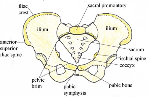
The anatomy of the female pelvis includes the pelvic bones, pelvic cavity, and surrounding muscles. The pelvic bones consist of the ilium, ischium, and pubis, which form the pelvic girdle. This girdle provides structural support and protection for pelvic organs.
The pelvic cavity houses important reproductive organs like the uterus, ovaries, and fallopian tubes. Muscles and ligaments within the pelvis assist in maintaining organ position and function. Understanding this anatomy is crucial for medical professionals in diagnosing and treating pelvic-related conditions. The female pelvis also plays a vital role in childbirth, accommodating and facilitating the passage of the baby through the birth canal.
Pelvic Structure
The female pelvis is a crucial part of the human body. It supports the weight of the upper body, allows movement, and plays a key role in childbirth. The pelvic structure is made up of bones, joints, and muscles that work together. Understanding the pelvic structure is important for health, especially for women.
Bone Composition
The pelvic bone structure consists of several key bones. These bones are strong and durable, providing support and protection.
- Ilium: This is the largest part of the pelvis. It forms the upper part and the sides.
- Ischium: This is the lower and back part of the pelvis. It supports the body’s weight when sitting.
- Pubis: This is the front part of the pelvis. It helps in forming the pelvic arch.
- Sacrum: This is a triangular bone at the base of the spine. It connects the spine to the pelvis.
- Coccyx: Known as the tailbone, it is the small, triangular bone at the very bottom of the spine.
Here is a table summarizing the bones:
| Bone | Location | Function |
|---|---|---|
| Ilium | Upper pelvis | Supports body weight |
| Ischium | Lower back pelvis | Supports while sitting |
| Pubis | Front pelvis | Forms pelvic arch |
| Sacrum | Base of spine | Spine to pelvis connection |
| Coccyx | Bottom of spine | Provides stability |
Joint Connections
The female pelvis has several important joint connections. These joints allow movement and flexibility.
- Sacroiliac Joint: This joint connects the sacrum to the ilium. It helps with weight transfer from the upper body to the legs.
- Pubic Symphysis: This joint is located at the front of the pelvis. It connects the left and right pubic bones. During childbirth, it can stretch to allow the baby to pass through.
- Hip Joints: These joints connect the pelvis to the femur (thigh bone). They allow leg movement and support the body’s weight.
Each joint has a specific role:
| Joint | Location | Function |
|---|---|---|
| Sacroiliac Joint | Between sacrum and ilium | Weight transfer |
| Pubic Symphysis | Front of pelvis | Connects pubic bones |
| Hip Joints | Between pelvis and femur | Leg movement |
The pelvic joints are vital for walking, sitting, and childbirth. They give the pelvis flexibility and strength.
Credit: www.open.edu
Pelvic Types
The anatomy of the female pelvis is fascinating and complex. The pelvis supports the weight of the upper body and protects the pelvic organs. Understanding the different pelvic types is essential for comprehending childbirth mechanics. Each type has unique characteristics that influence childbirth. The four main pelvic types are Gynecoid, Android, Anthropoid, and Platypelloid.
Gynecoid Shape
The Gynecoid shape is the most common type of female pelvis. It is considered the ideal shape for childbirth. This pelvic type has a round brim and wide pelvic cavity, providing ample space for the baby to pass through.
- Round brim: The brim is wide and circular.
- Wide pelvic cavity: The cavity is spacious, easing childbirth.
- Shallow depth: The pelvis is not very deep, making labor easier.
Here’s a table summarizing the Gynecoid pelvis characteristics:
| Feature | Description |
|---|---|
| Brim Shape | Round |
| Pelvic Cavity | Wide |
| Depth | Shallow |
The Gynecoid shape provides the best conditions for a smooth delivery. Its round brim and wide cavity offer the baby plenty of room to move through the birth canal. This pelvic type is most favorable for vaginal births.
Android Shape
The Android shape is more common in males but can also be found in females. It is characterized by a heart-shaped brim and a narrow pelvic cavity. This type can make childbirth more challenging.
- Heart-shaped brim: The brim is narrow and heart-shaped.
- Narrow pelvic cavity: The cavity is tight, restricting the baby’s passage.
- Deeper depth: The pelvis is deeper, complicating labor.
Here’s a table summarizing the Android pelvis characteristics:
| Feature | Description |
|---|---|
| Brim Shape | Heart-shaped |
| Pelvic Cavity | Narrow |
| Depth | Deeper |
The Android shape can make labor longer and more complicated. The narrow cavity and deeper depth limit the space for the baby. This type often results in a more difficult delivery.
Anthropoid Shape
The Anthropoid shape is oval in shape and is often found in women of African descent. It features a long anteroposterior diameter, making it unique among pelvic types.
- Oval brim: The brim is elongated and oval-shaped.
- Narrow transverse diameter: The width is narrow.
- Long anteroposterior diameter: The front-to-back measurement is long.
Here’s a table summarizing the Anthropoid pelvis characteristics:
| Feature | Description |
|---|---|
| Brim Shape | Oval |
| Pelvic Cavity | Narrow |
| Depth | Long anteroposterior |
The Anthropoid shape provides more room front-to-back but less room side-to-side. This can make certain birthing positions more effective. The long anteroposterior diameter can facilitate childbirth.
Platypelloid Shape
The Platypelloid shape is the least common of the pelvic types. It features a flat brim and a wide transverse diameter, making it quite distinct.
- Flat brim: The brim is flat and wide.
- Wide transverse diameter: The width is very wide.
- Shallow depth: The pelvis is not deep.
Here’s a table summarizing the Platypelloid pelvis characteristics:
| Feature | Description |
|---|---|
| Brim Shape | Flat |
| Pelvic Cavity | Wide transverse |
| Depth | Shallow |
The Platypelloid shape can complicate the engagement of the baby’s head during labor. The flat brim and wide transverse diameter pose unique challenges. This type often requires special attention during childbirth.
Muscle Layers
The female pelvis is a complex and fascinating part of the body. It supports many crucial functions. One important aspect is the muscle layers. These layers provide strength, stability, and support for pelvic organs. Understanding these muscles helps in recognizing their importance for health and movement.
Pelvic Floor Muscles
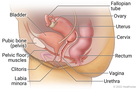
The pelvic floor muscles are a group of muscles located at the base of the pelvis. They play a key role in supporting the bladder, uterus, and bowel. These muscles also help in maintaining continence and providing stability during physical activities.
- Pubococcygeus: This muscle stretches from the pubic bone to the coccyx. It supports the pelvic organs and controls urine flow.
- Puborectalis: This muscle forms a sling around the rectum. It helps in maintaining fecal continence.
- Iliococcygeus: This muscle supports the pelvic organs and assists in lifting the pelvic floor.
A strong pelvic floor is crucial for various functions. Weakness in these muscles can lead to issues like urinary incontinence and pelvic organ prolapse. Exercises like Kegels can help strengthen them.
Core Stabilizers
The core stabilizers include several muscles that work together to provide stability to the pelvis and spine. These muscles are essential for maintaining posture and balance during movement.
- Transverse Abdominis: This deep muscle wraps around the abdomen. It acts like a natural corset, providing support to the spine.
- Multifidus: These small muscles run along the spine. They help in stabilizing and supporting the vertebrae during movement.
- Diaphragm: The main muscle for breathing. It also plays a role in core stability by maintaining intra-abdominal pressure.
A strong core is vital for everyday activities. It helps prevent back pain and improves overall physical performance. Engaging in exercises like planks and Pilates can enhance core strength.
Credit: myhealth.alberta.ca
Organs Within
The female pelvis houses a variety of important organs. These organs are crucial for reproductive health, urinary function, and overall well-being. Understanding the anatomy of the female pelvis can help in recognizing how these organs work together. Let’s explore the key organs within the female pelvis.
Reproductive Organs
The reproductive organs in the female pelvis are complex and essential for human life. They include the ovaries, fallopian tubes, uterus, and vagina.
- Ovaries: These small, almond-shaped glands produce eggs and hormones like estrogen and progesterone.
- Fallopian Tubes: These are narrow tubes that carry eggs from the ovaries to the uterus.
- Uterus: A pear-shaped organ where a fertilized egg implants and grows during pregnancy.
- Vagina: This muscular tube connects the cervix of the uterus to the outside of the body.
Each of these organs plays a vital role in reproduction. The ovaries release an egg each month during ovulation. The fallopian tubes then transport the egg. If fertilization occurs, it happens in the fallopian tubes. The fertilized egg travels to the uterus, where it may implant and grow. The vagina serves as the birth canal and also receives the penis during intercourse.
Here’s a table summarizing these organs:
| Organ | Function |
|---|---|
| Ovaries | Produce eggs and hormones |
| Fallopian Tubes | Transport eggs to the uterus |
| Uterus | Supports fetal development |
| Vagina | Birth canal and intercourse |
Urinary System
The urinary system in the female pelvis includes the bladder, urethra, and ureters. These organs work together to remove waste from the body.
- Bladder: A muscular sac that stores urine until it is ready to be expelled.
- Urethra: A tube that carries urine from the bladder to the outside of the body.
- Ureters: Two tubes that transport urine from the kidneys to the bladder.
The bladder holds urine until you feel the need to urinate. The urethra then allows the urine to leave the body. The ureters connect the kidneys to the bladder and continuously transport urine. These organs ensure that waste products are efficiently removed from the body. This helps maintain a healthy internal environment.
Here’s a simple breakdown of these organs:
| Organ | Function |
|---|---|
| Bladder | Stores urine |
| Urethra | Expels urine from the body |
| Ureters | Transport urine to the bladder |
Understanding these organs helps in recognizing how the body removes waste. Proper function of the urinary system is essential for overall health.
Blood Supply
The female pelvis is a fascinating and complex structure. Understanding its blood supply is crucial for medical professionals and students alike. The blood supply to the female pelvis ensures that all pelvic organs receive the oxygen and nutrients they need to function. Let’s delve deeper into the major arteries and venous drainage of the female pelvis.
Major Arteries
The blood supply to the female pelvis primarily comes from several major arteries. These arteries are essential for delivering oxygen-rich blood to the pelvic organs. The internal iliac artery is the main artery supplying the pelvis. It branches out into several smaller arteries:
- Uterine artery: Supplies blood to the uterus.
- Vaginal artery: Provides blood to the vagina.
- Obturator artery: Supplies the pelvic muscles.
- Internal pudendal artery: Feeds the perineum and external genitalia.
The internal iliac artery has two divisions:
- Anterior division: Supplies most of the pelvic organs.
- Posterior division: Supplies the gluteal region and sacrum.
Here is a table summarizing the major arteries and their branches:
| Artery | Branches | Supplied Areas |
|---|---|---|
| Internal Iliac Artery | Anterior & Posterior Divisions | Pelvic organs, gluteal region, sacrum |
| Uterine Artery | N/A | Uterus |
| Vaginal Artery | N/A | Vagina |
| Obturator Artery | N/A | Pelvic muscles |
| Internal Pudendal Artery | N/A | Perineum, external genitalia |
Venous Drainage
Venous drainage in the female pelvis is just as important as arterial supply. It ensures that deoxygenated blood is efficiently removed from the pelvic organs. The internal iliac vein is the primary vein responsible for this task. It collects blood from several smaller veins:
- Uterine veins: Drain blood from the uterus.
- Vaginal veins: Drain blood from the vagina.
- Obturator vein: Drains blood from pelvic muscles.
- Internal pudendal veins: Drain the perineum and external genitalia.
These veins converge into the internal iliac vein, which then merges with the common iliac vein. The common iliac vein eventually drains into the inferior vena cava, carrying blood back to the heart.
Here is a table summarizing the venous drainage:
| Vein | Drained Areas |
|---|---|
| Internal Iliac Vein | Pelvic organs, perineum, external genitalia |
| Uterine Veins | Uterus |
| Vaginal Veins | Vagina |
| Obturator Vein | Pelvic muscles |
| Internal Pudendal Veins | Perineum, external genitalia |
Nerve Innervation
The female pelvis is a complex structure that supports various vital functions. Understanding the anatomy involves delving into the nerve innervation that controls sensations, movements, and various bodily functions. Nerve innervation in the female pelvis is crucial for reproductive health, bladder control, and sensory perception.
Pelvic Nerves
The pelvic nerves are essential for transmitting signals between the brain and the pelvic region. These nerves include:
- Pudendal Nerve: This nerve is vital for controlling the external genitalia and the muscles of the pelvic floor.
- Hypogastric Nerve: It plays a role in the autonomic functions of the pelvis, including bladder and rectal control.
- Pelvic Splanchnic Nerves: These are responsible for the parasympathetic innervation of the pelvic organs.
The pudendal nerve, in particular, is responsible for sensory and motor functions. It splits into three branches:
- Inferior Rectal Nerve: Controls the anal sphincter.
- Perineal Nerve: Provides sensation to the perineum and controls perineal muscles.
- Dorsal Nerve of the Clitoris: Provides sensation to the clitoris.
The hypogastric nerve is part of the sympathetic nervous system and is responsible for:
- Contraction of the internal urethral sphincter.
- Contraction of the internal anal sphincter.
Pelvic splanchnic nerves originate from the sacral spinal cord and influence:
- Bladder contraction.
- Rectal motility.
- Sexual arousal.
Sensory Functions
Sensory functions of the pelvic nerves are critical for perception and response. These nerves help you feel sensations like touch, pain, and temperature.
The pudendal nerve is crucial for sensory functions in the pelvic region. It provides sensations to:
- The external genitalia.
- The perineum.
- The anus.
The hypogastric nerve contributes to the sensation of pelvic pain and discomfort. It helps in:
- Sensing bladder fullness.
- Detecting rectal filling.
Pelvic splanchnic nerves have a significant role in sexual arousal and orgasm. They affect:
- Clitoral engorgement.
- Vaginal lubrication.
Table of Sensory Functions:
| Nerve | Function |
|---|---|
| Pudendal Nerve | External genitalia sensation, perineal sensation, anal sensation |
| Hypogastric Nerve | Bladder fullness, rectal filling |
| Pelvic Splanchnic Nerves | Sexual arousal, vaginal lubrication |
In summary, nerve innervation in the female pelvis is vital for various functions. Proper understanding can help in diagnosing and treating pelvic disorders.
Pelvic Floor Function
The female pelvis is a complex structure with many important functions. One of its critical components is the pelvic floor. The pelvic floor is a group of muscles and tissues that stretch across the pelvis. It supports organs like the bladder, uterus, and rectum. Understanding the pelvic floor function helps in appreciating its role in everyday activities and health conditions.
Support Role
The pelvic floor acts as a support system for the pelvic organs. These organs include the bladder, uterus, and rectum. Without the pelvic floor, these organs would not stay in place. This support role is crucial for maintaining normal bodily functions.
Here are some key functions:
- Supports bladder: Prevents urinary incontinence.
- Supports uterus: Helps in menstrual function and reproductive health.
- Supports rectum: Aids in bowel control.
These functions ensure that the organs work correctly. The pelvic floor muscles also help in stabilizing the spine and maintaining posture. This stabilization is vital for movements and physical activities.
A strong pelvic floor is essential for activities like:
- Lifting heavy objects
- Exercising
- Maintaining balance
Here is a table summarizing the support roles:
| Organ | Support Function |
|---|---|
| Bladder | Prevents incontinence |
| Uterus | Supports reproductive health |
| Rectum | Aids in bowel control |
Childbirth Impact
Childbirth significantly impacts the pelvic floor. During delivery, the pelvic floor muscles stretch to allow the baby to pass through. This stretching can cause weakness or damage to the muscles.
Here are some common impacts:
- Pelvic floor weakness: Leads to urinary incontinence.
- Pelvic organ prolapse: Organs may drop from their normal position.
- Chronic pelvic pain: Caused by muscle strain or injury.
After childbirth, many women experience pelvic floor dysfunction. This dysfunction can affect daily activities and quality of life. Pelvic floor exercises, also known as Kegels, can help in recovery. These exercises strengthen the muscles and improve support functions.
Here are some benefits of pelvic floor exercises:
- Improves bladder control
- Reduces risk of prolapse
- Enhances sexual health
Timely intervention and exercises can restore pelvic floor health. It’s important to consult healthcare providers for proper guidance.
Credit: childrenswi.org
Clinical Relevance
The anatomy of the female pelvis is crucial for understanding various medical conditions and treatments. Clinical relevance highlights the importance of this anatomical region in diagnosing and managing pelvic disorders. It also guides surgical interventions to ensure patient safety and successful outcomes.
Pelvic Disorders
Pelvic disorders can significantly impact a woman’s quality of life. Understanding the anatomy of the female pelvis helps in diagnosing and treating these conditions effectively.
- Pelvic Inflammatory Disease (PID): This infection affects the female reproductive organs and can lead to severe complications if not treated promptly. Symptoms include pelvic pain, fever, and abnormal vaginal discharge.
- Endometriosis: This condition occurs when tissue similar to the lining inside the uterus grows outside it. It causes chronic pain and can lead to infertility.
- Pelvic Organ Prolapse: This happens when pelvic organs drop from their normal position due to weakened support structures. Symptoms include a feeling of heaviness in the pelvis and urinary issues.
Surgical Considerations
Understanding the anatomy of the female pelvis is essential for surgical planning and execution. Surgeons must navigate this complex region with precision to avoid complications.
- Hysterectomy: Removing the uterus requires a thorough knowledge of pelvic anatomy to avoid damage to surrounding organs and tissues.
- Pelvic Reconstructive Surgery: This surgery corrects pelvic organ prolapse. Surgeons must understand the pelvic floor’s anatomy to restore proper function.
- Cesarean Section: During this procedure, the surgeon must carefully navigate the pelvic region to deliver the baby safely while minimizing risks to the mother.
The table below summarizes important structures and their clinical significance:
| Structure | Clinical Significance |
|---|---|
| Uterus | Central to reproductive health; involved in conditions like fibroids and cancer |
| Ovaries | Important for hormone production; affected by cysts and cancer |
| Pelvic Floor Muscles | Support pelvic organs; involved in prolapse and incontinence |
References/further Reading
The female pelvis is a complex structure that plays a crucial role in various bodily functions, including childbirth. Understanding its anatomy is essential for medical professionals, students, and anyone interested in human biology. For those who wish to delve deeper into the subject, this section provides a comprehensive list of references and further reading materials.
Citations:
To ensure the accuracy of the information presented, it is important to rely on reliable and peer-reviewed sources. Here are some key citations related to the anatomy of the female pelvis:
- Gray’s Anatomy – This classic medical textbook offers detailed descriptions and illustrations of the female pelvis.
- Journal of Obstetrics and Gynaecology – This journal publishes research papers on various aspects of obstetrics and gynecology, including pelvic anatomy.
- Netter’s Atlas of Human Anatomy – This atlas provides high-quality anatomical illustrations, making it easier to understand the complex structures of the pelvis.
Below is a table summarizing key resources:
| Resource | Description |
|---|---|
| Gray’s Anatomy | Comprehensive medical textbook with detailed descriptions |
| Journal of Obstetrics and Gynaecology | Peer-reviewed journal with research papers on pelvic anatomy |
| Netter’s Atlas of Human Anatomy | Illustrative atlas for easier understanding of anatomical structures |
Further Reading:
For those who wish to expand their knowledge beyond the basics, the following materials offer in-depth information and insights:
- Williams Obstetrics – This textbook provides comprehensive coverage of obstetrics, including detailed chapters on pelvic anatomy.
- Clinical Anatomy by Regions – This book breaks down anatomical information by regions, offering detailed insights into the pelvis.
- Pelvic Floor Disorders: Imaging and Multidisciplinary Approach to Management – This book focuses on the clinical aspects of pelvic floor disorders, with detailed anatomical descriptions.
These resources are invaluable for medical students, healthcare professionals, and researchers:
- Williams Obstetrics – Known for its detailed and comprehensive approach to obstetrics.
- Clinical Anatomy by Regions – Offers a regional approach to anatomy for easier understanding.
- Pelvic Floor Disorders – Focuses on the clinical management of pelvic floor disorders with anatomical insights.
Frequently Asked Questions
What Is The Normal Anatomy Of The Female Pelvis?
The female pelvis includes the hip bones, sacrum, and coccyx. It houses the bladder, uterus, ovaries, and rectum. The pelvic floor muscles provide support.
What Is The Pelvic Pain Line Anatomy?
The pelvic pain line separates visceral pain regions of the pelvis. It helps differentiate pain originating above or below the line.
What Are The 4 Types Of Female Pelvis?
The four types of female pelvis are gynecoid, android, anthropoid, and platypelloid. Gynecoid is the most common and favorable for childbirth. Android resembles a male pelvis. Anthropoid is elongated, and platypelloid is flat and wide.
What Is The Part Of The Pelvis That Sticks Out?
The part of the pelvis that sticks out is called the iliac crest. It is the top edge of the hip bone.
Conclusion
Understanding the female pelvis is crucial for many medical fields. This knowledge helps in childbirth, surgeries, and diagnosing conditions. By studying its anatomy, healthcare providers can offer better care. Keep learning to stay informed and healthy. The female pelvis is a fascinating and vital part of human anatomy.


