 Reproductive Health Sexual and Reproductive Health

Gynecological anatomy involves the study of the female reproductive system, including organs like the ovaries, uterus, and vagina. It plays a crucial role in women’s health.
Gynecological anatomy is the foundation of understanding female reproductive health. The primary organs include the ovaries, which produce eggs, and the uterus, where a fertilized egg implants and grows. The vagina acts as the birth canal and the exit for menstrual flow.
The fallopian tubes connect the ovaries to the uterus, facilitating egg transport. These organs work together to support reproductive functions and hormonal balance. Proper knowledge of gynecological anatomy aids in diagnosing and treating various health conditions, promoting overall well-being. Understanding this complex system is essential for medical professionals and women alike.
Female Reproductive System
The female reproductive system is a complex and fascinating part of gynecological anatomy. It consists of several organs, each playing a crucial role in reproduction. Understanding these organs and their functions can help in maintaining reproductive health. Let’s dive into the major organs and their functionalities.
Major Organs
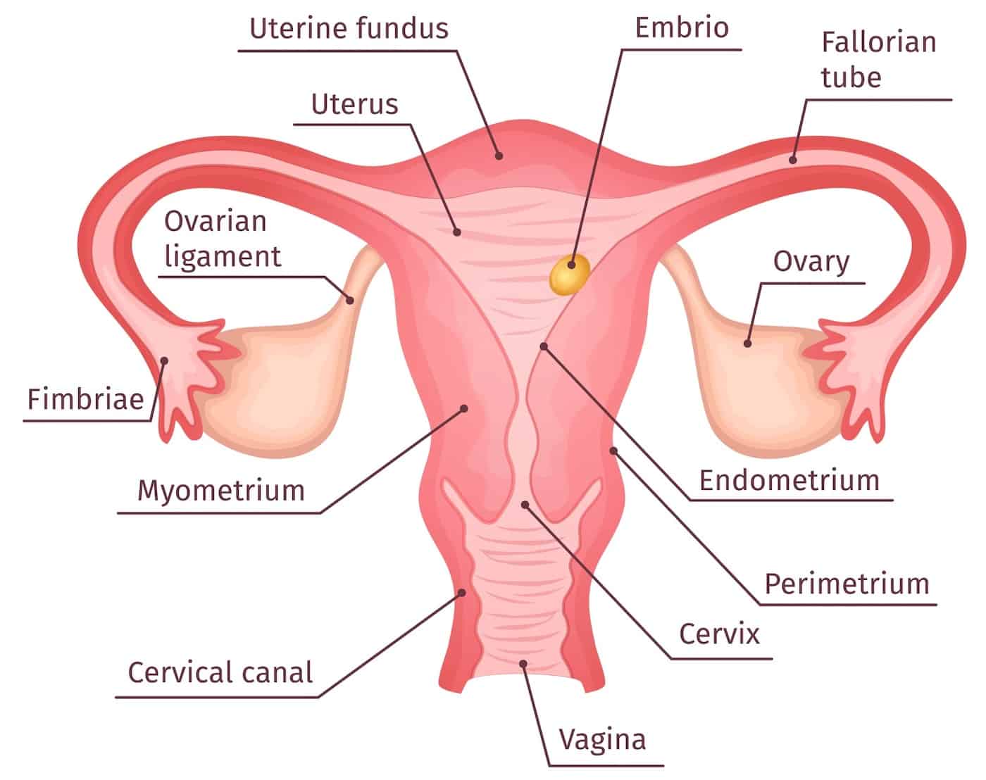
The female reproductive system includes several key organs. Each of these organs has a unique role in the reproductive process. Here are the primary organs involved:
- Ovaries: Two small, oval-shaped organs located on either side of the uterus. They produce eggs and hormones such as estrogen and progesterone.
- Fallopian Tubes: These are narrow tubes that connect the ovaries to the uterus. They are the pathway for the eggs to travel from the ovaries to the uterus.
- Uterus: A hollow, pear-shaped organ where a fertilized egg implants and grows during pregnancy. The uterus has a thick muscular wall that can expand and contract.
- Cervix: The lower part of the uterus that opens into the vagina. It acts as a gateway between the uterus and the vagina.
- Vagina: A muscular canal that connects the cervix to the external body. It serves as the birth canal and also allows for menstrual flow to exit the body.
Below is a table summarizing the major organs and their primary functions:
| Organ | Primary Function |
|---|---|
| Ovaries | Produce eggs and hormones |
| Fallopian Tubes | Transport eggs to the uterus |
| Uterus | House and nourish a fertilized egg |
| Cervix | Connect uterus to the vagina |
| Vagina | Serve as the birth canal |
Credit: anatomywarehouse.com
Functionality
The functionality of the female reproductive system is intricate and essential for reproduction. Let’s explore how these organs work together:
Ovaries: The ovaries release eggs during ovulation, which occurs roughly once a month. They also produce hormones that regulate the menstrual cycle and support pregnancy.
Fallopian Tubes: During ovulation, an egg travels from the ovary through the fallopian tube. Fertilization by sperm usually happens here. The fertilized egg then moves to the uterus.
Uterus: The uterus prepares for a potential pregnancy each month. Its lining thickens to support a fertilized egg. If fertilization occurs, the egg implants in the uterine wall and begins to grow. If not, the lining sheds during menstruation.
Cervix: The cervix produces mucus that changes in consistency during the menstrual cycle. This mucus can help or hinder sperm movement. During childbirth, the cervix dilates to allow the baby to pass through.
Vagina: The vagina receives sperm during intercourse. It also serves as the passageway for menstrual blood and the baby during childbirth.
Understanding these functions helps in recognizing the importance of reproductive health and the vital role each organ plays in the process of conception and childbirth.
Ovaries
Gynecological anatomy is a complex field that focuses on the female reproductive system. The ovaries play a vital role in this system. These small, almond-shaped organs are crucial for reproductive health and hormone production. Understanding their structure and function is key to appreciating their importance.
Structure
The ovaries are located on each side of the uterus in the lower abdomen. Each ovary is about the size of an almond. They are held in place by ligaments attached to the uterus. The structure of an ovary includes:
- Outer Cortex: This layer contains follicles, which house immature eggs.
- Inner Medulla: This area is rich in blood vessels, nerves, and connective tissue.
- Follicles: Each follicle holds a single egg and goes through stages of development.
A table summarizing the parts of an ovary:
| Part | Description |
|---|---|
| Outer Cortex | Houses follicles and immature eggs |
| Inner Medulla | Contains blood vessels and nerves |
| Follicles | Develop and release eggs |
Each ovary takes turns releasing an egg every month. This process is called ovulation. The structure of the ovary supports this function by providing a nurturing environment for the egg to mature.
Hormonal Role
The ovaries are essential for hormone production. They produce estrogen and progesterone, which are key female hormones. These hormones regulate the menstrual cycle and support pregnancy. The hormonal role of the ovaries includes:
- Estrogen Production: Estrogen helps develop female secondary sexual characteristics. It also regulates the menstrual cycle.
- Progesterone Production: Progesterone prepares the uterus for a fertilized egg. It maintains the early stages of pregnancy.
A table summarizing the hormones produced by the ovaries:
| Hormone | Function |
|---|---|
| Estrogen | Develops female characteristics and regulates the menstrual cycle |
| Progesterone | Prepares the uterus for pregnancy and supports early pregnancy |
Hormonal balance is crucial for reproductive health. Imbalances can cause menstrual irregularities, fertility issues, and other health problems. The ovaries work closely with the pituitary gland and hypothalamus to regulate hormone levels.
Fallopian Tubes
The fallopian tubes are a crucial part of the female reproductive system. These narrow tubes connect the ovaries to the uterus. They play a key role in the process of fertilization and pregnancy. Understanding the anatomy and function of the fallopian tubes can provide insight into how the reproductive system works.
Anatomy
The fallopian tubes are about 10-12 cm long and have several parts:
- Fimbriae: These finger-like projections help in capturing the egg released from the ovary.
- Infundibulum: This funnel-shaped part lies next to the fimbriae.
- Ampulla: The widest section, where fertilization typically occurs.
- Isthmus: A narrow section that connects to the uterus.
- Intramural part: The segment that passes through the uterine wall.
The fallopian tubes are lined with ciliated cells. These cells have tiny hair-like structures. They help move the egg toward the uterus. The walls of the tubes have smooth muscles. These muscles contract to assist the egg’s movement.
| Part | Function |
|---|---|
| Fimbriae | Capture the egg |
| Infundibulum | Funnels the egg |
| Ampulla | Site of fertilization |
| Isthmus | Narrow passage to uterus |
| Intramural part | Passes through uterine wall |
Fertilization Process
The fertilization process often begins in the ampulla of the fallopian tubes. Here’s a simple breakdown:
- Ovulation: The ovary releases an egg.
- Fimbriae capture the egg and guide it into the infundibulum.
- Cilia and muscle contractions move the egg through the fallopian tube.
- Sperm travel up the fallopian tube to meet the egg.
- Fertilization usually occurs in the ampulla. A sperm fuses with the egg, forming a zygote.
- The zygote travels to the uterus over several days. It continues to divide and grow.
- Implantation: The zygote implants itself in the uterine lining, starting pregnancy.
This journey is crucial for successful fertilization and pregnancy. Any blockage or damage to the fallopian tubes can prevent this process. Regular check-ups can help ensure the fallopian tubes are healthy and functional.
:max_bytes(150000):strip_icc():format(webp)/female-reproductive-system-with-image-diagram-538949875-96ab73c46e5b45f0ba6ff254d1609a83.jpg)
Credit: www.verywellhealth.com
Uterus
The uterus, a vital organ in the female reproductive system, plays a key role in pregnancy, childbirth, and the menstrual cycle. Located in the pelvis, it resembles an inverted pear and serves as the nurturing environment for a developing fetus. Understanding the structure and function of the uterus is essential for comprehending many aspects of gynecological health.
Layers Of The Uterus
The uterus has three distinct layers, each with specific functions:
- Endometrium: This is the innermost layer. It thickens during the menstrual cycle to prepare for a potential pregnancy. If fertilization does not occur, the endometrium sheds, leading to menstruation.
- Myometrium: The middle layer consists of smooth muscle. It contracts during menstruation and childbirth. These contractions help expel menstrual blood and push the baby out during labor.
- Perimetrium: This is the outermost layer. It is a thin layer of tissue that covers the uterus. It provides structural support and protection.
The following table summarizes the layers of the uterus and their functions:
| Layer | Location | Function |
|---|---|---|
| Endometrium | Innermost | Thickens for pregnancy, sheds during menstruation |
| Myometrium | Middle | Contracts for menstruation and childbirth |
| Perimetrium | Outermost | Provides structural support and protection |
Menstrual Cycle
The menstrual cycle is a monthly process that prepares the female body for pregnancy. It involves several stages:
- Menstrual Phase: This is the start of the cycle. The endometrium sheds, leading to menstrual bleeding. It lasts about 3-7 days.
- Follicular Phase: This phase begins after menstruation. The endometrium thickens again. Follicles in the ovaries mature, preparing for ovulation.
- Ovulation: This occurs midway through the cycle. A mature egg is released from the ovary. It travels down the fallopian tube, ready for fertilization.
- Luteal Phase: This phase follows ovulation. If the egg is fertilized, it implants in the endometrium. If not, the endometrium prepares to shed again.
Hormones like estrogen and progesterone regulate the menstrual cycle. They ensure the endometrium thickens and sheds at the right times. Irregularities in the cycle can indicate health issues. Keeping track of the menstrual cycle helps in identifying such issues early.
Cervix
The cervix is an essential part of the female reproductive system. It acts as the gateway between the vagina and the uterus. Understanding the cervix’s structure and functions provides insight into many aspects of women’s health. This blog section focuses on the cervix’s anatomical features and its role in reproduction.
Anatomical Features
The cervix is a cylindrical-shaped organ located at the lower part of the uterus. It measures about 2-3 centimeters in length. The cervix consists of two main parts:
- Endocervix: The inner part that connects to the uterus.
- Exocervix: The outer part that extends into the vagina.
The cervical canal runs through the cervix, allowing passage between the uterus and vagina. The cervical canal has two openings:
- Internal os: The opening towards the uterus.
- External os: The opening towards the vagina.
The cervix is made up of fibrous tissue, collagen, and blood vessels. Its surface is covered by two types of cells:
- Squamous cells: Found on the exocervix.
- Glandular cells: Found on the endocervix.
These features allow the cervix to perform its functions effectively. Regular check-ups, including Pap smears, help in early detection of any abnormalities in the cervix.
Role In Reproduction
The cervix plays a crucial role in reproduction. During the menstrual cycle, the cervix undergoes changes to facilitate or prevent the passage of sperm. During ovulation, the cervix produces mucus that is thin and slippery, making it easier for sperm to travel through the cervical canal.
In pregnancy, the cervix changes to protect the developing baby. It produces a thick mucus plug that seals the cervical canal, preventing infections. This mucus plug remains until labor.
During labor, the cervix dilates and effaces (thins out) to allow the baby to pass through the birth canal. The degree of dilation is measured in centimeters, from 0 (closed) to 10 (fully dilated). Effacement is measured in percentages, from 0% (not effaced) to 100% (fully effaced).
The table below summarizes the cervical changes during pregnancy and labor:
| Stage | Cervical Changes |
|---|---|
| Pregnancy | Formation of mucus plug |
| Early Labor | Beginning of dilation and effacement |
| Active Labor | Full dilation (10 cm) and effacement (100%) |
Understanding the cervix’s role in reproduction highlights its importance in women’s health. Regular gynecological care is vital for monitoring cervical health and ensuring successful pregnancies.

Credit: www.aboutcancer.com
Vagina
Understanding gynecological anatomy is essential for women’s health. One of the key components of this anatomy is the vagina. The vagina is a muscular tube that plays a crucial role in the female reproductive system. It connects the external genitals to the uterus, allowing for a range of functions vital to reproductive health.
Structure And Length
The structure of the vagina is quite unique. It is a muscular canal that extends from the vulva to the cervix. The walls of the vagina are lined with mucous membranes which keep it moist and elastic. This allows it to stretch during intercourse and childbirth.
On average, the vagina measures about 3 to 4 inches in length when not aroused. During sexual arousal, it can expand and lengthen to accommodate penetration. This feature is due to its muscular and elastic nature.
Here is a summary table for a better understanding:
| Aspect | Details |
|---|---|
| Length (at rest) | 3 to 4 inches |
| Length (aroused) | Can expand longer |
| Lining | Mucous membranes |
| Function | Elastic and muscular |
The vagina is also rich in blood vessels and nerves, making it sensitive to touch. The upper part of the vagina is more sensitive to pressure, while the lower part is more sensitive to touch. This distribution plays a role in sexual pleasure and reproductive health.
Functions
The vagina has several important functions in the female reproductive system. One of its primary roles is to serve as the passageway for menstrual flow. During menstruation, blood and tissue from the uterus exit the body through the vagina.
Another critical function is its role in sexual intercourse. The vagina allows for the penetration of the penis, which is essential for conception. Sperm deposited in the vagina can travel through the cervix and into the uterus to fertilize an egg.
Here are some additional functions:
- Childbirth: The vagina serves as the birth canal through which a baby is delivered.
- Self-cleaning: The vagina has natural secretions that help keep it clean and maintain a healthy pH balance.
- Protection: The acidic environment of the vagina helps protect against infections by preventing the growth of harmful bacteria.
The vagina also plays a role in sexual pleasure, thanks to its sensitivity and ability to expand. This makes it an essential part of the sexual experience for many women.
Understanding the structure and functions of the vagina helps in appreciating its role in reproductive health. Regular gynecological check-ups ensure it remains healthy and functions properly.
External Genitalia
Gynecological anatomy is a fascinating subject that helps us understand the female reproductive system. The external genitalia are the outer parts of this system. These parts play key roles in protection, sensation, and sexual function. Knowing about them helps in better health and well-being.
Labia
The labia are important parts of the external genitalia. They come in two sets: the labia majora and the labia minora.
Labia Majora:
- Also called the “outer lips”.
- They are larger and contain fat tissue.
- They protect the inner parts of the genitalia.
- Covered with pubic hair after puberty.
Labia Minora:
- Also called the “inner lips”.
- They are smaller and more delicate.
- They are hairless and have many nerve endings.
- They provide protection to the vaginal and urethral openings.
Both sets of labia have different colors and sizes. They vary from person to person. The labia minora are rich in blood vessels, making them very sensitive. The labia majora cover and protect the inner parts from injury and infections.
Clitoris
The clitoris is a small but very sensitive part of the external genitalia. It’s located at the top of the labia minora.
Key Features of the Clitoris:
- It’s about the size of a pea.
- Contains thousands of nerve endings.
- Main function is to provide sexual pleasure.
- Covered by the clitoral hood, which protects it.
The clitoris has a small visible part and a larger internal structure. The internal part extends into the body. It swells and becomes more sensitive during arousal. The clitoris is the only human organ solely for pleasure.
Understanding the clitoris helps in better sexual health. It’s important to know that it can be different in size and shape. Everyone is unique, and that’s normal.
Pelvic Floor
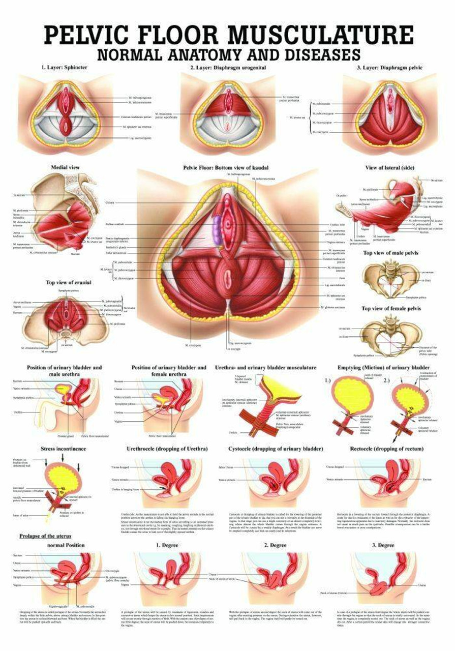
The pelvic floor is a key part of gynecological anatomy. It supports many organs in the lower abdomen, including the bladder, uterus, and rectum. Understanding the pelvic floor’s structure can help maintain good health and prevent issues.
Muscles Involved
The pelvic floor consists of several important muscles. These muscles form a hammock-like structure that supports pelvic organs. Key muscles include:
- Levator ani: This group includes the pubococcygeus, iliococcygeus, and puborectalis muscles.
- Coccygeus: Also known as the ischiococcygeus, this muscle supports the tailbone.
- Perineal muscles: These muscles are located between the anus and the genitals.
Each muscle plays a unique role. The levator ani muscles are the main support for pelvic organs. They help with urination, bowel movements, and childbirth. The coccygeus muscle supports the tailbone and helps stabilize the spine. The perineal muscles assist in controlling the openings of the urethra and anus.
Weak or damaged pelvic floor muscles can lead to problems. Common issues include urinary incontinence, pelvic organ prolapse, and difficulty with bowel movements. Strengthening these muscles through exercises like Kegels can help maintain their function.
Importance For Health
The pelvic floor’s health is crucial for overall well-being. A strong pelvic floor ensures proper support for pelvic organs. This support helps prevent pelvic organ prolapse, where organs like the bladder or uterus fall out of place. A healthy pelvic floor also aids in controlling bladder and bowel functions.
Maintaining a strong pelvic floor can help during pregnancy and childbirth. During pregnancy, the pelvic floor supports the growing baby. A strong pelvic floor can make childbirth easier and reduce the risk of complications. Postpartum, strong muscles help in recovery and prevent long-term issues.
Here are some benefits of a healthy pelvic floor:
- Prevents urinary incontinence
- Reduces the risk of pelvic organ prolapse
- Enhances sexual function and satisfaction
- Supports bowel control
Regular exercises, such as Kegels, can strengthen the pelvic floor. It is important to practice these exercises correctly to avoid strain. Consulting with a healthcare provider can provide guidance and ensure proper technique.
Credit: wfoflou.com
References/further Reading
Gynecological Anatomy is a complex and vital aspect of women’s health. Understanding it is crucial for both medical professionals and patients. To dive deeper into this topic, References/Further Reading provides essential resources and research papers that offer in-depth knowledge and insights.
Citations:
Here are some key citations that can help you better understand gynecological anatomy:
- Williams Gynecology, 4th Edition: A comprehensive textbook covering all aspects of gynecology, including anatomy, pathology, and treatment.
- Gray’s Anatomy for Students: This book offers detailed anatomical descriptions and is essential for medical students.
- ACOG Practice Bulletin: The American College of Obstetricians and Gynecologists publishes guidelines and practice bulletins that are invaluable for clinical practice.
- Journal of Obstetrics and Gynecology: A peer-reviewed journal featuring the latest research and reviews in the field.
For those interested in specific anatomical structures, PubMed offers a plethora of research articles. Here’s a quick table summarizing some of the most cited works:
| Title | Author(s) | Year | Journal |
|---|---|---|---|
| Anatomy of the Female Pelvis | Smith et al. | 2018 | Journal of Anatomy |
| Histology of the Uterus | Jones & Brown | 2020 | Histology Reviews |
Further Reading:
To expand your knowledge, consider exploring the following resources:
- Netter’s Atlas of Human Anatomy: This atlas offers detailed illustrations that are perfect for visual learners.
- Clinical Gynecologic Endocrinology and Infertility: This book provides a deep dive into the hormonal and reproductive aspects of gynecology.
- UpToDate: An evidence-based clinical resource that offers comprehensive information on various gynecological conditions.
- MedlinePlus: A reliable source for patient-friendly information about gynecological health.
For more specialized topics, consider these journals and websites:
- Human Reproduction: Focuses on reproductive biology and gynecology.
- Gynecologic Oncology: Covers cancers affecting the female reproductive system.
- WebMD: Offers articles and guides on a wide range of gynecological issues.
These resources provide a solid foundation for anyone looking to delve deeper into the anatomy and physiology of the female reproductive system. Happy reading!
Frequently Asked Questions
What Is Gynecological Anatomy?
Gynecological anatomy refers to the structure of the female reproductive system. It includes organs like the ovaries, fallopian tubes, uterus, and vagina. Understanding this anatomy is crucial for diagnosing and treating various conditions.
Why Is Gynecological Anatomy Important?
Gynecological anatomy is important for diagnosing and treating female reproductive health issues. It helps medical professionals understand how organs function and interact. This knowledge aids in surgeries, fertility treatments, and disease management.
What Organs Are In The Female Reproductive System?
The female reproductive system includes the ovaries, fallopian tubes, uterus, and vagina. These organs work together to support reproduction and menstrual cycles. Each organ has a specific function essential for overall reproductive health.
How Does The Uterus Function?
The uterus is a muscular organ where a fertilized egg implants and grows. It supports fetal development during pregnancy. The uterine lining sheds during menstruation if no pregnancy occurs.
Conclusion
Understanding gynecological anatomy is vital for women’s health. Knowledge empowers individuals to make informed healthcare decisions. Regular check-ups and awareness can prevent many issues. Always consult with a healthcare professional for personalized advice. Stay informed and proactive about your gynecological health for a better quality of life.


