Reproductive Health Sexual and Reproductive Health
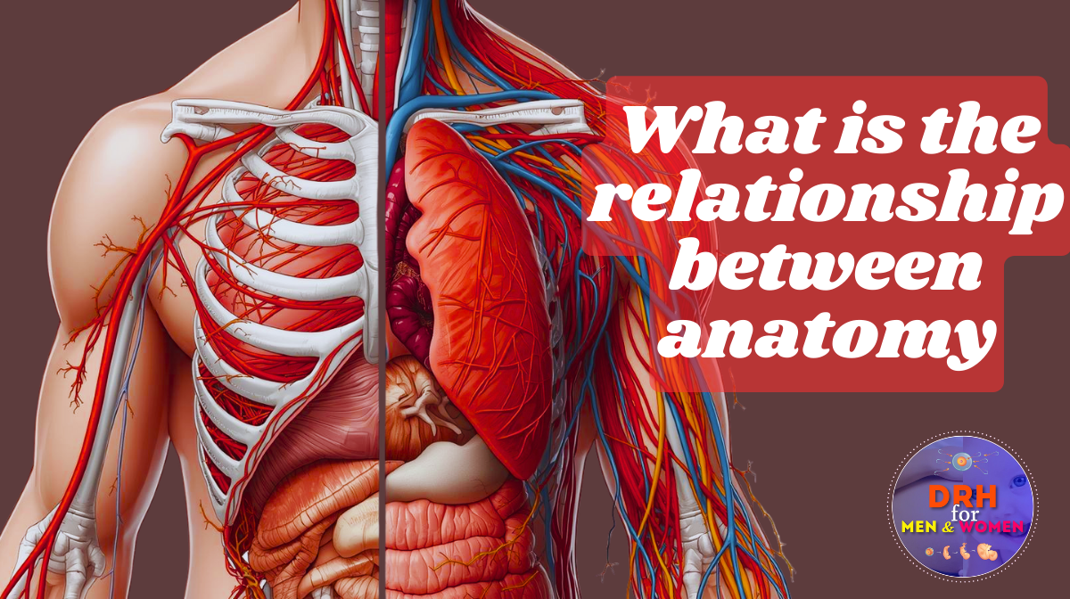 Anatomy Explained: The Blueprint of the Human Body
The human body is often compared to a complex machine, but even the most advanced supercomputer or luxury car pales in comparison to biological engineering. We breathe without thinking, heal from cuts automatically, and process complex thoughts in milliseconds. But to understand how this machine runs, we first have to understand the parts that build it. This is the domain of anatomy.
Anatomy is the scientific study of the structure of organisms and their parts. Derived from the Greek words ana (up) and tome (cutting), it literally means “to cut up.” While the definition sounds simple, the field is vast. It serves as the foundation for all medical knowledge. Without a map of the body’s terrain, doctors, surgeons, and physical therapists would be navigating blindly.
Whether you are a medical student, a fitness enthusiast, or simply curious about how you tick, understanding anatomy offers a profound appreciation for the vessel you live in every day.
From Sketches to Scanners: A Brief History
The study of anatomy isn’t new. It has been a central part of human curiosity for millennia. Ancient Egyptians gained a rudimentary understanding of organs through the mummification process, though their knowledge was more religious than scientific.
The Greeks, particularly Galen in the 2nd century AD, advanced the field significantly, although much of his work was based on animal dissection and contained errors that persisted for centuries. It wasn’t until the Renaissance that human anatomy took a giant leap forward. Andreas Vesalius, often called the father of modern anatomy, challenged Galen’s long-standing theories by performing public dissections on human cadavers. His meticulous drawings in De Humani Corporis Fabrica (On the Fabric of the Human Body) set a new standard for accuracy.
Today, we have moved far beyond simple sketches. While cadaver dissection remains a rite of passage for medical students, technology has revolutionized how we see inside ourselves, moving from hand-drawn diagrams to high-definition digital rendering.
The Three Levels of Anatomical Study
Because the body is so complex, anatomists break the study down into manageable levels, ranging from what you can see with the naked eye to structures so small they require powerful magnification.
Gross Anatomy
When people hear the word “anatomy,” this is usually what they picture. Gross anatomy (or macroscopic anatomy) deals with structures large enough to be seen without a microscope. This includes the heart, lungs, bones, and muscles.
There are generally two ways to approach gross anatomy:
- Systemic Anatomy: This method studies the body by systems. For example, you might study the entire skeletal system (osteology) or the entire muscular system (myology) at once, regardless of where the parts are located.
- Regional Anatomy: This approach focuses on specific areas. For instance, a medical student might study the “head and neck” region, examining every bone, muscle, nerve, and blood vessel in that specific area before moving on to the thorax or the leg.
Microscopic Anatomy (Histology)
Life happens at the cellular level. Microscopic anatomy, also known as histology, dives into structures that are invisible to the naked eye. By examining thin slices of tissue under a microscope, scientists can see the organization of cells that make up our organs.
Understanding histology is vital because disease often starts here. Cancer, for example, is a disruption of cellular function. By understanding what normal lung tissue looks like under a microscope, pathologists can identify the abnormal cell growth that characterizes a tumor.
Developmental Anatomy (Embryology)
Our bodies are not static; they change dramatically over time. Developmental anatomy traces structural changes that occur in the body throughout the lifespan. Embryology is a specialized branch of this field that focuses on developmental changes that occur before birth.
Studying embryology explains the logic behind some of our strange anatomical quirks. For example, the reason nerves sometimes take long, winding paths through the body is often because of how the embryo folded and grew during the first few weeks of development.
Anatomy vs. Physiology: Structure Meets Function
It is impossible to fully understand anatomy without discussing its partner: physiology. If anatomy is the study of the body’s structure (the “what” and “where”), physiology is the study of how those parts work (the “how” and “why”).
The two are inextricably linked by the principle of complementarity of structure and function. This means that what a body part is dictates what it does.
Consider the bones. They are made of hard mineral deposits, which allows them to protect organs and support body weight. If bones were soft, they couldn’t perform this function. Conversely, the lungs are composed of incredibly thin tissue. This structural thinness allows oxygen to pass quickly into the bloodstream. If lungs were thick and hard like bone, breathing would be impossible.
In essence, anatomy provides the map, and physiology explains the traffic flowing through the roads. You cannot master one without a working knowledge of the other.
Why It Matters: Clinical Significance
Anatomy is not just an academic exercise; it is the cornerstone of clinical practice. Every diagnosis, surgical procedure, and physical therapy session relies on anatomical knowledge.
- Physical Examinations: When a doctor presses on your abdomen (palpation), they are visualizing the anatomy beneath your skin. They know exactly where the liver ends and the stomach begins. If they feel a mass where it shouldn’t be, anatomical knowledge tells them which organ is likely affected.
- Surgical Precision: In surgery, millimeters matter. A surgeon must know the precise location of nerves and blood vessels to repair a hernia or remove an appendix without causing collateral damage.
- Medical Imaging: Radiologists rely on anatomy to interpret X-rays and CT scans. Recognizing the difference between a normal anatomical shadow and a fracture or tumor is a skill built entirely on understanding structure.
The Future: Modern Techniques and Virtual Reality
The study of anatomy is currently undergoing a digital renaissance. While traditional dissection is still valuable, it is now supplemented by incredible technology.
Non-invasive imaging techniques like Magnetic Resonance Imaging (MRI) and Computed Tomography (CT) scans allow us to view the living body in cross-sections without making a single incision. Even more exciting is the rise of 3D modeling and virtual reality (VR).
Medical students can now put on VR headsets and “walk through” the human heart or practice surgical procedures on a virtual patient that can be reset with the click of a button. These tools make anatomy more accessible and interactive, allowing for a deeper understanding of spatial relationships that textbooks simply cannot convey.
The Never-Ending Study
Anatomy is often viewed as a “complete” science—after all, we have counted every bone and named every muscle. However, our understanding of the body continues to evolve. We are still discovering new nuances in how tissues interact, how fascia connects the body in continuous webs, and how individual anatomical variations affect health.
From the microscopic cells that power our metabolism to the structural framework that allows us to stand upright, anatomy is the story of us. It is the blueprint of human life, and the more we study it, the more we can appreciate the sophisticated, resilient, and beautiful design of the human body.