 Reproductive Health Sexual and Reproductive Health

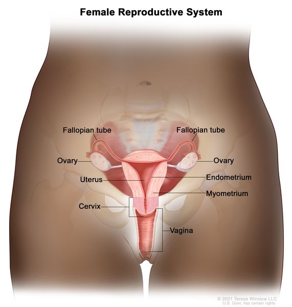
Female reproductive anatomy includes the ovaries, fallopian tubes, uterus, and vagina. These organs work together for reproduction.
The female reproductive system plays a crucial role in human reproduction. The ovaries produce eggs and hormones. The fallopian tubes transport eggs from the ovaries to the uterus. The uterus provides a nurturing environment for a fertilized egg to develop into a fetus.
The vagina serves as the birth canal and allows for the passage of menstrual flow. This complex system also regulates the menstrual cycle and hormonal balance. Understanding female reproductive anatomy is essential for maintaining reproductive health and addressing potential issues. Proper care and regular check-ups can help identify and prevent reproductive health problems.
External Anatomy
The female reproductive system is a complex and intricate network of organs. It plays a crucial role in human reproduction. The external anatomy of the female reproductive system includes several important structures. These external parts are vital for various functions including protection, sensation, and sexual activity.
Vulva
The vulva is the external part of the female genitalia. It includes the mons pubis, labia majora, labia minora, clitoris, and the openings to the urethra and vagina. The vulva serves multiple functions such as protecting the internal genital organs and providing sexual pleasure.
Key components of the vulva:
- Mons pubis: Fatty tissue covering the pubic bone.
- Labia majora: Outer lips that protect the inner structures.
- Labia minora: Inner lips that surround the vaginal and urethral openings.
- Clitoris: A small, sensitive organ providing sexual pleasure.
The vulva is rich in nerve endings, making it highly sensitive to touch. Understanding the anatomy of the vulva is essential for sexual health and hygiene.
Clitoris
The clitoris is a small, sensitive organ located at the top of the vulva. It is often referred to as the center of female sexual pleasure. The clitoris has thousands of nerve endings, making it extremely sensitive.
Parts of the clitoris include:
- Glans: The visible, external part of the clitoris.
- Shaft: The body of the clitoris, which extends internally.
- Crura: Two “legs” that extend downwards along the pubic bones.
The primary function of the clitoris is to provide sexual pleasure. Stimulation of the clitoris can lead to sexual arousal and orgasm. It is important to recognize the clitoris’ role in female sexual health.
Labia
The labia are part of the vulva and consist of the labia majora and labia minora. These structures protect the internal genital organs and contribute to sexual sensation.
Labia majora:
- These are the larger, outer lips.
- They contain fatty tissue and sweat glands.
- They protect the inner parts of the vulva.
Labia minora:
- These are the smaller, inner lips.
- They are located inside the labia majora.
- They surround the openings to the vagina and urethra.
- They are rich in blood vessels and nerve endings.
The labia help in protecting the vaginal and urethral openings. They also play a role in sexual arousal and lubrication. Understanding the labia’s anatomy is vital for maintaining sexual health and hygiene.

Credit: www.healthdirect.gov.au
Internal Structures
The female reproductive system is a marvel of biological engineering. It consists of both external and internal structures. The internal structures play vital roles in reproduction and overall health. Let’s explore these internal structures in more detail.
Vagina
The vagina is a muscular tube connecting the external genitals to the uterus. It serves multiple roles in the female reproductive system. It is the passage through which menstrual blood leaves the body. The vagina also acts as the birth canal during childbirth. It allows for sexual intercourse.
Here are some key features of the vagina:
- Elasticity: The vagina can stretch to accommodate intercourse and childbirth.
- Self-cleaning: The vagina maintains a healthy environment through natural secretions.
- Microbiome: Healthy bacteria in the vagina help prevent infections.
The vaginal wall has several layers:
| Layer | Description |
|---|---|
| Mucosa | Innermost layer that produces lubrication. |
| Muscularis | Middle layer of smooth muscle. |
| Adventitia | Outer layer providing structural support. |
The vagina’s health is crucial for overall reproductive health. Regular check-ups and good hygiene can help maintain a healthy vagina.
Cervix
The cervix is a small, cylindrical structure that connects the vagina to the uterus. It serves as a gateway between the lower and upper reproductive tracts. The cervix has two main parts: the endocervix and the ectocervix.
The endocervix is the inner part of the cervix. It contains a canal that opens into the uterus. The ectocervix is the outer part that extends into the vagina. The cervix has several important functions:
- Barrier: The cervix acts as a barrier to protect the uterus from infections.
- Menstrual flow: It allows menstrual blood to flow from the uterus to the vagina.
- Pregnancy: The cervix changes during pregnancy to help keep the fetus in place.
The cervix also produces mucus. This mucus changes in consistency during the menstrual cycle. It becomes thinner around ovulation, aiding sperm movement into the uterus.
Regular Pap smears are crucial. They help detect changes in cervical cells that could indicate cancer.
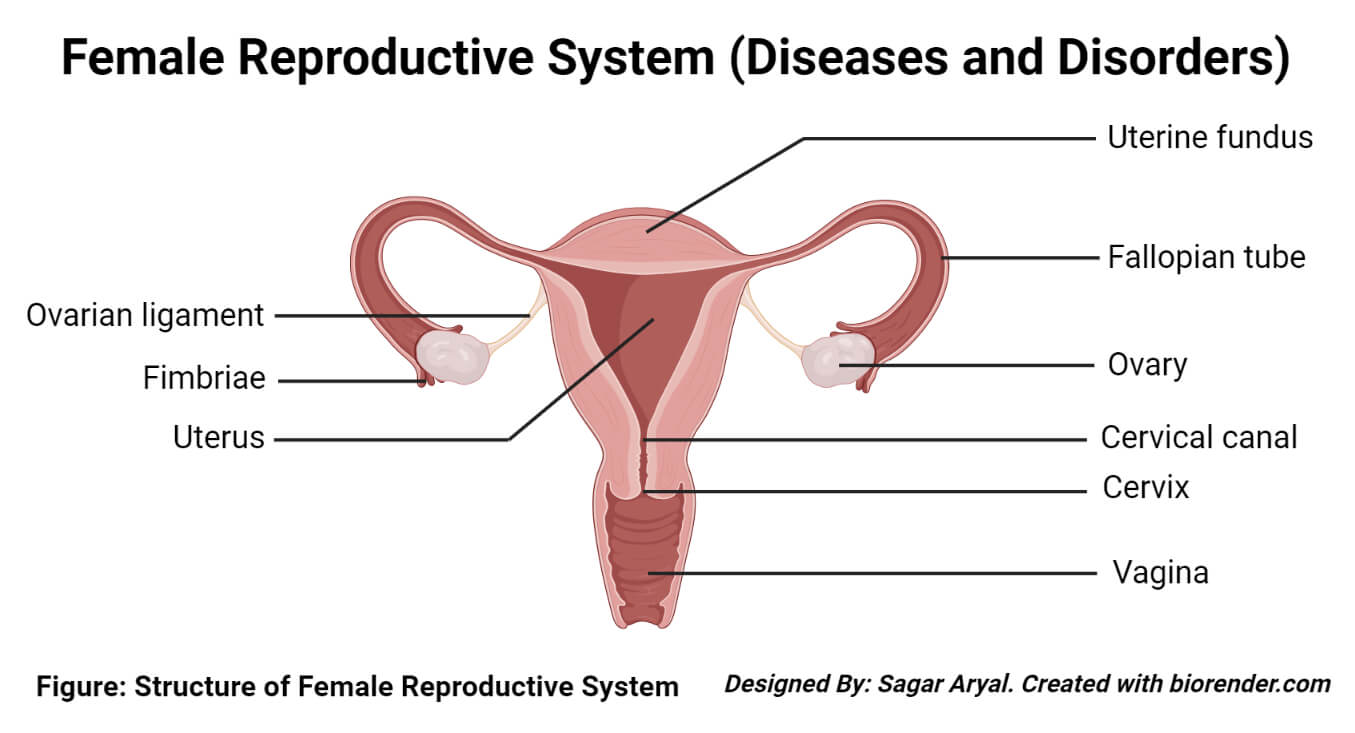
Uterus
The uterus is a pear-shaped organ located in the pelvis. It plays a central role in reproduction. The uterus has three main parts: the fundus, body, and cervix.
The fundus is the top part of the uterus. The body is the main part. The cervix connects the uterus to the vagina. The uterus has several important functions:
- Menstruation: The uterus sheds its lining during menstruation.
- Pregnancy: The uterus supports the developing fetus during pregnancy.
- Labor: The uterus contracts during labor to help deliver the baby.
The uterine wall has three layers:
| Layer | Description |
|---|---|
| Endometrium | Inner lining that thickens and sheds during the menstrual cycle. |
| Myometrium | Middle muscular layer that contracts during childbirth. |
| Perimetrium | Outer protective layer. |
The health of the uterus is vital for fertility and overall reproductive health. Conditions like fibroids and endometriosis can affect the uterus. Regular medical check-ups can help monitor and maintain uterine health.

Credit: www.medicalnewstoday.com
Ovaries
The female reproductive system is a marvel of nature. At the heart of this system are the ovaries. These small, almond-shaped organs play a crucial role in reproduction. They are located on either side of the uterus. The ovaries have two main functions: they produce eggs and secrete hormones.
Function
The primary function of the ovaries is to produce and release eggs. This process is known as ovulation. Each month, one ovary releases a mature egg. This egg travels down the fallopian tube where it may meet sperm for fertilization.
Ovaries also have a crucial role in maintaining the health of the female reproductive system. They do this by:
- Producing eggs (ova) which are essential for reproduction.
- Regulating the menstrual cycle by releasing eggs in a cyclical pattern.
- Supporting pregnancy by producing hormones that sustain the uterine lining.
Below is a table summarizing the key functions of the ovaries:
| Function | Description |
|---|---|
| Egg Production | Producing ova for fertilization. |
| Menstrual Cycle Regulation | Controlling the monthly cycle by releasing eggs. |
| Hormone Secretion | Producing hormones that regulate reproductive health. |
Hormone Production
The ovaries are also responsible for producing vital hormones. These hormones play a key role in female health. The main hormones produced by the ovaries are estrogen and progesterone.
Estrogen helps develop female characteristics. It regulates the menstrual cycle. It also maintains the health of the reproductive organs.
Progesterone prepares the uterus for pregnancy. It thickens the uterine lining. This helps support a fertilized egg. If the egg is not fertilized, progesterone levels drop, leading to menstruation.
Here is a brief overview of the hormones produced by the ovaries:
- Estrogen: Develops and maintains female characteristics, regulates menstrual cycle.
- Progesterone: Prepares the uterus for pregnancy, supports early pregnancy.
The balance of these hormones is crucial. It affects not just reproductive health but overall well-being.
Credit: www.cancer.gov
Fallopian Tubes
The female reproductive system is a complex and amazing part of the human body. One of its key components is the fallopian tubes. These tubes play a crucial role in reproduction. They transport eggs from the ovaries to the uterus. Let’s dive into the details of the fallopian tubes.
Structure
The fallopian tubes are narrow tubes. Each tube measures about 10 to 12 cm in length. They are located on each side of the uterus. The tubes connect the ovaries to the uterus. The structure of the fallopian tubes can be divided into four main parts:
- Infundibulum: The funnel-shaped end near the ovary. It has finger-like projections called fimbriae.
- Ampulla: The widest section of the tube. This is where fertilization usually occurs.
- Isthmus: The narrow section near the uterus.
- Interstitial (or Intramural): The part that passes through the uterine wall.
The inside of the fallopian tubes is lined with cilia. These tiny hair-like structures help move the egg towards the uterus. The tube’s muscular walls also contract to assist this movement. The fallopian tubes are vital for transporting the egg and sperm.
Role In Fertilization
The fallopian tubes play a crucial role in fertilization. After ovulation, the egg is released from the ovary. The fimbriae of the infundibulum catch the egg. The egg then moves into the ampulla.
During sexual intercourse, sperm travels through the female reproductive tract. The sperm reaches the ampulla of the fallopian tube. Here, one sperm fertilizes the egg. This process is called fertilization.
Once fertilization occurs, the fertilized egg, now called a zygote, travels down the tube. The cilia and muscular contractions help move the zygote towards the uterus. In the uterus, the zygote implants into the uterine lining and begins to grow into a baby.
The fallopian tubes are essential for this journey. Without them, the egg and sperm could not meet. Fertilization and pregnancy would not be possible. Thus, fallopian tubes are a vital part of female reproductive anatomy.
Menstrual Cycle
The menstrual cycle is an essential aspect of female reproductive anatomy. It is a natural process that prepares the body for potential pregnancy. The cycle involves a series of changes in the ovaries and the lining of the uterus. Understanding the menstrual cycle is crucial for women’s health and wellness. It typically lasts around 28 days, but this can vary from person to person.
Phases
The menstrual cycle is divided into four main phases. Each phase plays a unique role in preparing the body for pregnancy. These phases are:
- Menstrual Phase: This phase marks the beginning of the cycle. It involves the shedding of the uterine lining. Women experience bleeding during this phase, which lasts 3-7 days.
- Follicular Phase: This phase starts on the first day of menstruation and continues until ovulation. The pituitary gland releases follicle-stimulating hormone (FSH), which stimulates the ovaries to produce follicles. One of these follicles will mature into an egg.
- Ovulation Phase: Ovulation occurs around the middle of the cycle. The mature egg is released from the ovary. This phase is the most fertile time in the cycle.
- Luteal Phase: After ovulation, the luteal phase begins. The ruptured follicle forms a structure called the corpus luteum, which secretes hormones to thicken the uterine lining.
A clear understanding of these phases can help in tracking menstrual health. It also aids in recognizing any irregularities in the cycle.
Hormonal Changes
Hormonal changes are crucial to the menstrual cycle. Different hormones rise and fall during the cycle, affecting the body in various ways. The key hormones involved include:
| Hormone | Function |
|---|---|
| Estrogen | Stimulates the growth of the uterine lining during the follicular phase. |
| Follicle-Stimulating Hormone (FSH) | Promotes the growth of ovarian follicles. |
| Luteinizing Hormone (LH) | Triggers ovulation and the release of the mature egg. |
| Progesterone | Maintains the thickened uterine lining during the luteal phase. |
During the menstrual phase, levels of estrogen and progesterone are low. This triggers the shedding of the uterine lining. In the follicular phase, FSH levels rise, leading to the growth of follicles. Estrogen levels also increase, thickening the uterine lining. During the ovulation phase, a spike in LH causes the release of the egg. In the luteal phase, progesterone levels rise, ensuring the uterine lining is ready for a possible pregnancy.
These hormonal changes are crucial for the proper functioning of the menstrual cycle. They regulate the different phases and prepare the body for potential pregnancy.
Pregnancy Changes
The female reproductive anatomy undergoes significant transformations during pregnancy. These changes help support the growing baby and prepare the mother’s body for childbirth. Understanding these changes is essential for expecting mothers and those supporting them.
Anatomical Adaptations
During pregnancy, several anatomical adaptations occur to accommodate the developing fetus. These changes affect various parts of the female reproductive system:
- Uterus: The uterus expands significantly. It starts about the size of a fist and grows to accommodate the baby, placenta, and amniotic fluid.
- Cervix: The cervix softens and dilates. This process, called effacement, prepares the cervix for the baby’s passage during delivery.
- Breasts: The breasts grow larger and more tender. This happens to prepare for milk production and breastfeeding.
Below is a table summarizing the key anatomical changes:
| Body Part | Change |
|---|---|
| Uterus | Expands from fist-size to accommodate baby |
| Cervix | Softens and dilates |
| Breasts | Grow larger and more tender |
Physiological Changes
Pregnancy also brings about several physiological changes in a woman’s body. These changes are crucial for the health of both the mother and the baby:
- Hormonal Shifts: Hormone levels, such as estrogen and progesterone, increase significantly. These hormones help maintain the pregnancy and prepare the body for labor.
- Blood Volume: The blood volume increases by 30-50%. This increase supports the growing fetus and placenta.
- Metabolic Rate: The metabolic rate rises. This helps meet the energy demands of pregnancy.
Expecting mothers might also experience changes in skin pigmentation, increased urination, and digestive adjustments. These physiological changes are normal and indicate a healthy pregnancy.
In summary, both anatomical and physiological changes are vital for a successful pregnancy. These transformations help ensure the baby’s growth and prepare the mother’s body for childbirth.

Credit: www.nursinghero.com
Common Disorders
The female reproductive system is complex and fascinating. It plays a crucial role in the continuation of life. Despite its importance, it is prone to certain disorders. These common disorders can affect women’s health and their quality of life. Understanding these disorders helps in early detection and treatment. Let’s explore two prevalent conditions: Endometriosis and Fibroids.
Endometriosis
Endometriosis is a painful disorder. Tissue similar to the lining inside the uterus grows outside it. This tissue can appear on the ovaries, fallopian tubes, and other pelvic organs. It causes severe pain, especially during periods.
Symptoms of endometriosis include:
- Pelvic pain and cramping
- Heavy menstrual bleeding
- Pain during intercourse
- Infertility
Diagnosis of endometriosis usually involves:
- Pelvic exam
- Ultrasound
- MRI
- Laparoscopy
Treatment options for endometriosis vary. They depend on the severity and symptoms. Common treatments include:
- Pain medications
- Hormone therapy
- Surgical options to remove endometrial tissue
Early diagnosis and treatment can improve the quality of life.
Fibroids
Fibroids are non-cancerous growths in the uterus. They vary in size and number. Many women with fibroids experience no symptoms. For those who do, symptoms can range from mild to severe.
Common symptoms of fibroids include:
- Heavy menstrual bleeding
- Pelvic pain and pressure
- Frequent urination
- Constipation
- Backache
Fibroids are often discovered during routine pelvic exams. Diagnosis may involve:
- Ultrasound
- MRI
- Hysteroscopy
Treatment for fibroids depends on symptoms and size. Options include:
- Medications to regulate hormones
- Non-invasive procedures like MRI-guided focused ultrasound
- Surgical options like myomectomy or hysterectomy
Managing fibroids can significantly enhance women’s well-being.
Health And Wellness
The female reproductive anatomy is a complex and vital part of women’s health. Understanding how to maintain health and wellness in this area is crucial. Proper care and attention can prevent many health issues and improve overall well-being. This section covers the importance of regular check-ups and the role of nutrition and lifestyle in female reproductive health.
Regular Check-ups
Regular check-ups are essential for maintaining female reproductive health. Visiting a gynecologist annually helps detect issues early. Early detection can prevent serious conditions and ensure timely treatment.
Key benefits of regular check-ups include:
- Early detection of diseases like ovarian cysts, fibroids, and cancers.
- Monitoring menstrual health to identify irregularities.
- Screening for sexually transmitted infections (STIs) to maintain sexual health.
During a check-up, doctors perform various tests:
| Test | Purpose |
|---|---|
| Pap Smear | Detects cervical cancer and precancerous changes. |
| Pelvic Exam | Checks for abnormalities in the reproductive organs. |
| HPV Test | Identifies the presence of the human papillomavirus. |
It’s important to discuss any symptoms or concerns with your doctor. This open communication ensures you receive the best care and guidance.
Nutrition And Lifestyle
Nutrition and lifestyle choices play a significant role in reproductive health. A balanced diet and healthy habits can improve overall well-being and prevent many health issues.
Key nutrients for reproductive health:
- Folic Acid: Essential for DNA synthesis and repair, and crucial during pregnancy.
- Iron: Prevents anemia and supports healthy blood flow.
- Calcium and Vitamin D: Important for bone health, especially during menopause.
Healthy lifestyle habits include:
- Regular exercise: Helps regulate hormones and maintain a healthy weight.
- Avoiding smoking and excessive alcohol: Reduces the risk of reproductive health issues.
- Managing stress: Lowers the risk of menstrual irregularities and other health problems.
Creating a balanced diet and incorporating exercise into your routine can have significant benefits. Consider consulting a nutritionist for personalized advice.
Credit: microbenotes.com
References/further Reading
The female reproductive system is a complex and fascinating subject. Learning about it helps us understand how life begins and how our bodies function. For those interested in delving deeper, we’ve compiled a list of reliable sources and additional readings. These resources will further enhance your knowledge and appreciation of female reproductive anatomy.
Citations:
Understanding the female reproductive anatomy requires accurate and credible information. Here are some key sources that provide detailed and authoritative insights:
- “Human Anatomy & Physiology” by Elaine N. Marieb and Katja Hoehn: This textbook is widely used in medical and health science courses. It offers an in-depth look at the reproductive system, complete with diagrams and explanations.
- “Gray’s Anatomy” by Henry Gray: A classic reference, this book offers detailed descriptions and illustrations of the human body. It includes a comprehensive section on the female reproductive system.
- National Institutes of Health (NIH): The NIH provides extensive online resources about reproductive health, including anatomy, function, and common disorders. Visit their website for up-to-date and peer-reviewed information.
The following table summarizes these citations:
| Source | Description |
|---|---|
| “Human Anatomy & Physiology” | Comprehensive textbook with diagrams and explanations. |
| “Gray’s Anatomy” | Classic reference with detailed descriptions and illustrations. |
| NIH | Online resources with peer-reviewed information. |
Further Reading:
For those eager to explore more about the female reproductive system, here are some additional readings that provide valuable insights:
- “Our Bodies, Ourselves” by the Boston Women’s Health Book Collective: This book is an essential guide to women’s health and sexuality, including a detailed section on reproductive anatomy and health.
- “The Vagina Bible” by Dr. Jen Gunter: This book addresses common myths and misconceptions about female anatomy and reproductive health, providing factual and empowering information.
- Planned Parenthood: Their website offers comprehensive guides and articles on various aspects of reproductive health, including anatomy, contraception, and menstrual health.
These resources are excellent for anyone seeking a deeper understanding of the female reproductive system. They offer both scientific perspectives and practical health advice.
Remember, knowledge about our bodies empowers us to make informed health decisions. Dive into these readings to broaden your understanding and appreciation of female reproductive anatomy.
Frequently Asked Questions
What Are The Main Parts Of The Female Reproductive System?
The main parts include the ovaries, fallopian tubes, uterus, and vagina. Each part has a specific function in reproduction.
How Do Ovaries Function In Reproduction?
Ovaries produce eggs and hormones like estrogen and progesterone. These hormones regulate the menstrual cycle and pregnancy.
What Is The Role Of Fallopian Tubes?
Fallopian tubes transport eggs from the ovaries to the uterus. Fertilization usually occurs in the fallopian tubes.
What Happens In The Uterus?
The uterus nurtures a fertilized egg, allowing it to develop into a fetus. It is essential for pregnancy.
Conclusion
Understanding female reproductive anatomy is vital for overall health. This knowledge empowers women to make informed health decisions. Regular check-ups can help detect any issues early. Always consult healthcare professionals for accurate information. Share this knowledge to promote awareness and well-being among women.
Stay informed, stay healthy.


